The aging of bones is a complex process. As the body ages, the composition and mechanical properties of bones change. Newer research is targeting pathways that regulate these changes. The following are some of the factors known to affect bone aging. Then, learn how to protect your bones as you get older. Several simple lifestyle changes can protect you against bone aging. You’ll also learn how to take care of your bones and keep them strong.
Vitamin-D levels
Low vitamin-D levels have been linked to reduced mineralization of bone and a reduced bone mass. This may explain why people who have low vitamin-D levels are more susceptible to bone fractures. Researchers led by Robert Ritchie at the University of California, Berkeley, Materials Science, and Engineering Department, and Berkeley Lab’s Materials Sciences Division conducted this study. These findings highlight the important role of vitamin D in aging and bone health.

Decreased vitamin-D levels prevent bone remodeling and slow bone re-mineralization, two processes that are required to maintain healthy bones throughout life. The study also measured bone quality on a 100-micrometer scale, the diameter of a human hair. The researchers monitored cracks and the amount of resistance to fractures over time in the participants’ bones. The researchers concluded that vitamin-D deficiency inhibits bone remodeling and may lead to early bone aging.
Research has indicated that inadequate absorption of calcium may also negatively affect bone health. Intestinal absorption of calcium is regulated by vitamin D in the form of 1,25-dihydroxy vitamin D3 (also known as vitamin D3). Ionized calcium and parathyroid hormone also regulate calcium homeostasis. Therefore, the current review focuses on the role of vitamin D in calcium homeostasis and changes related to aging.
Glucocorticoids
Glucocorticoids regulate the lineage commitment of mesenchymal progenitors, or osteoblasts, which are the main precursors of bone. Glucocorticoids are known to promote osteoblastogenesis by stimulating the production of Wnt and sFRP1. Glucocorticoids also increase b-catenin expression and decrease PPARg levels, which are key factors in the formation of osteoblasts.
Glucocorticoids have both anabolic and catabolic effects on bone. The catabolic actions of glucocorticoids are believed to be due to their ability to induce the production of Wnt proteins by osteoblasts. This biphasic effect of glucocorticoids on bone may be a cause of its disparate effects. In addition, glucocorticoids regulate Wnt inhibitors in mature osteoblasts, resulting in decreased bone density and reduced bone mass.
During the inflammatory process, endogenous glucocorticoids may overwhelm the bone-forming process. This may be one of the reasons for age-related osteoporosis and inflammation-associated osteoporosis. However, the exact mechanisms of bone loss and resorption are not understood. However, the role of endogenous glucocorticoids in bone development is unclear.
Cellular senescence
As we age, we experience cellular changes that impair fracture healing. In particular, osteogenic cells become more senescent and exhibit impaired proliferation and functional activity. These changes are particularly evident in osteocytes. One approach to reverse this process is the treatment of aged rats with LMV, which inhibits osteogenic cell senescence through the Sirt1/p53/p21 axis. It works by stimulating osteogenic cell proliferation and inhibiting the senescent response in mice, which increases bone formation and strengthens bones in aged rats.
The vibration treatment was effective in improving osteoblast functions. The results also showed that vibration treatment improved the expression of p21 and decreased DNA damage in osteoblasts. The researchers are now investigating how vibration can influence cellular senescence in other tissues. They hope to eventually develop a treatment for osteoporosis, one of the most common causes of bone aging. The effects of vibration on the aging of bones should be studied in humans.
In mice, cellular senescence is a key regulator of postnatal bone development. Targeting cellular senescence is one way to prevent bone development retardation in female offspring after PDE. The study also shows the potential for antenatal corticosteroid therapy in pregnant women. The treatment could help them achieve a healthy pregnancy. But it is not yet clear if antenatal corticosteroid therapy could prevent or reduce the risk of bone senescence.
Melatonin
The relationship between melatonin and the aging of bones is complicated, but it may be as simple as a sleep hormone. The hormone acts on the osteoclast gene, which regulates bone formation. It also regulates MT2R, an enzyme that controls osteoblast differentiation. Several studies have linked melatonin to increase bone mass. Researchers have mapped the role of melatonin in osteoporosis, and the effects of melatonin on bone formation and microarchitecture.

The antioxidant enzyme SOD2 is also involved in osteogenesis. Melatonin has been shown to enhance a molecule called vascular endothelial growth factor (VEGF), which contributes to bone formation. Moreover, melatonin treatment elevates VEGF during the formation of granulation tissue and accelerates the angiogenic process. In this way, melatonin protects the bones against inflammatory bone disease.
The cellular components in the bone microenvironment include osteoblasts and osteoclasts. Each plays a critical role in the bone turnover cycle. Bone aging results in many bone-related diseases, which can have a detrimental effect on one’s quality of life. In the case of aging of bones, melatonin’s role in bone metabolism is intriguing. In addition, the hormone inhibits osteoclastogenesis, increases osteoblast-induced expression of osteoprotegerins, and suppresses bone metastasis.
Telomere length
Telomeres, which are highly conserved repeats of nucleotides, are responsible for preventing the deterioration of double-stranded DNA ends in cells. Studies have linked the shortening of telomeres to the development of age-related diseases. Telomere length decreases with each round of cell division, causing cellular aging. It is also implicated in the aging of the bones, as telomere shortening limits the growth and differentiation of bone cells.
Despite this apparent paradox, the relationship between telomere length and bone health has gained increasing attention in recent years. A study by Kveiborg et al. analyzed the relationship between telomere length and bone mineral density in septuagenarians and elderly people. Telomere length negatively correlated with age and BMD. However, the findings of the study were not statistically significant. Furthermore, the authors noted that telomere length was not associated with incident fractures or BMD changes over the course of seven years.
The study also suggests that people with shortened telomeres may be at a higher risk for developing diseases such as cancer. Scientists may be able to prevent cancer cells by blocking telomerase, but this would impair fertility, wound healing, and the production of blood cells and immune system cells. Geneticist Richard Cawthon has studied the relationship between telomere length and the aging of bones.
Mechanisms
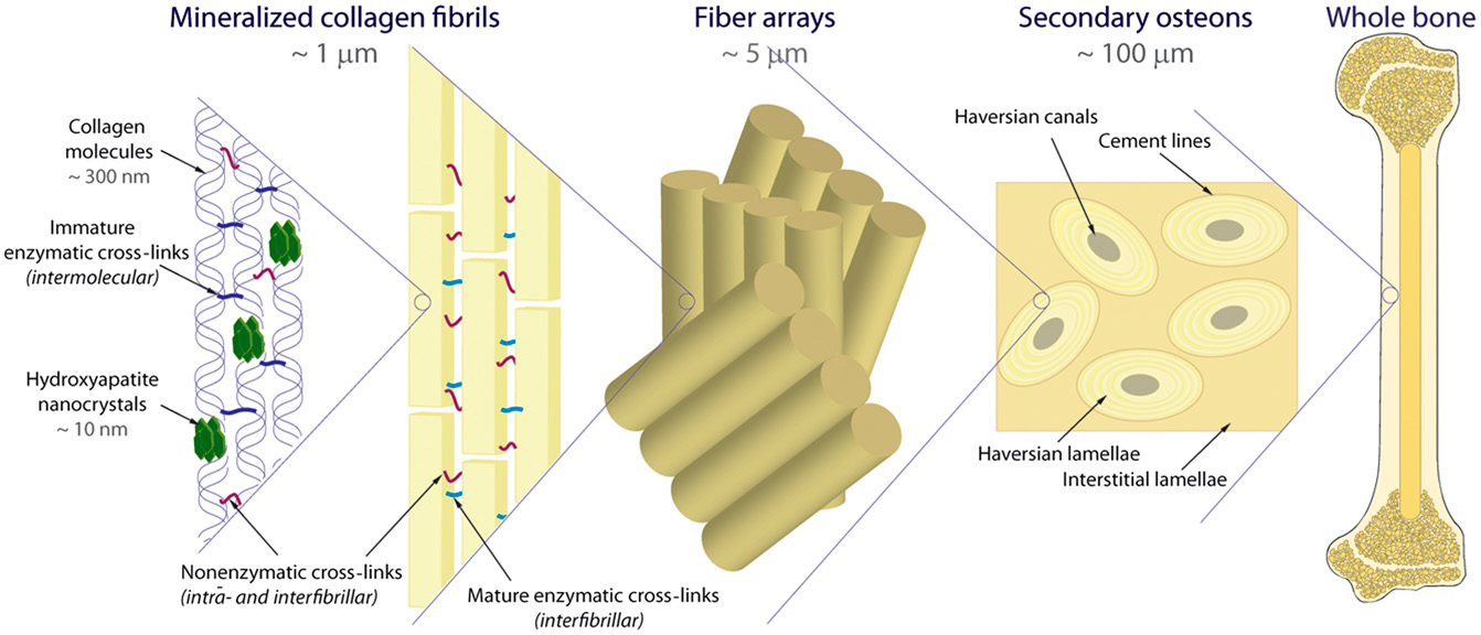
Molecular mechanisms of bone aging are important for our pursuit of a longer life. While long-lived bone cells are most vulnerable to the damaging effects of aging, alterations in the environment also affect short-lived cells. Therefore, a better understanding of the pro and anti-aging molecules and signaling pathways within aged bone is critical. By better understanding these networks, we can develop therapeutic strategies for age-related osteoporosis.
Changing cellular environments are known to cause degeneration in bone. In addition, the aging environment is altered to favor short-lived cells. Changes in endogenous glucocorticoids, oxidized lipids, and sex steroids may contribute to the degeneration of bone. This article summarizes current knowledge about aging and bone. Emphasis will be placed on the role of ROS and autophagy in bone aging.
Cells with an altered secretome are considered senescent. This phenotype may serve a paracrine, autocrine, or endocrine function. This review will discuss pathways leading to a senescence profile in the aged skeleton, as well as the implications for age-related osteoporosis and its treatment. Cellular senescence may also be a target for new therapeutic options.
Prevention
The bones are the foundation of our body, providing structure and protection to our organs. They also store calcium and phosphorus. As we age, our bones lose density and become more brittle. The good news is that there are several steps you can take to avoid osteoporosis and keep your bones strong and healthy. Here are some of these measures. Ensure you get plenty of calcium and other nutrients from your diet.

The first step to prevention is to understand the risk factors for osteoporosis. Women are more susceptible to this condition than men, and it’s estimated that one in every two women and one in four men will suffer from osteoporosis at some point in their lives. While osteoporosis is an inherited trait of women, it can be prevented or delayed by following the appropriate preventive measures. To avoid this disease, you need to understand its causes, symptoms, and possible treatments.
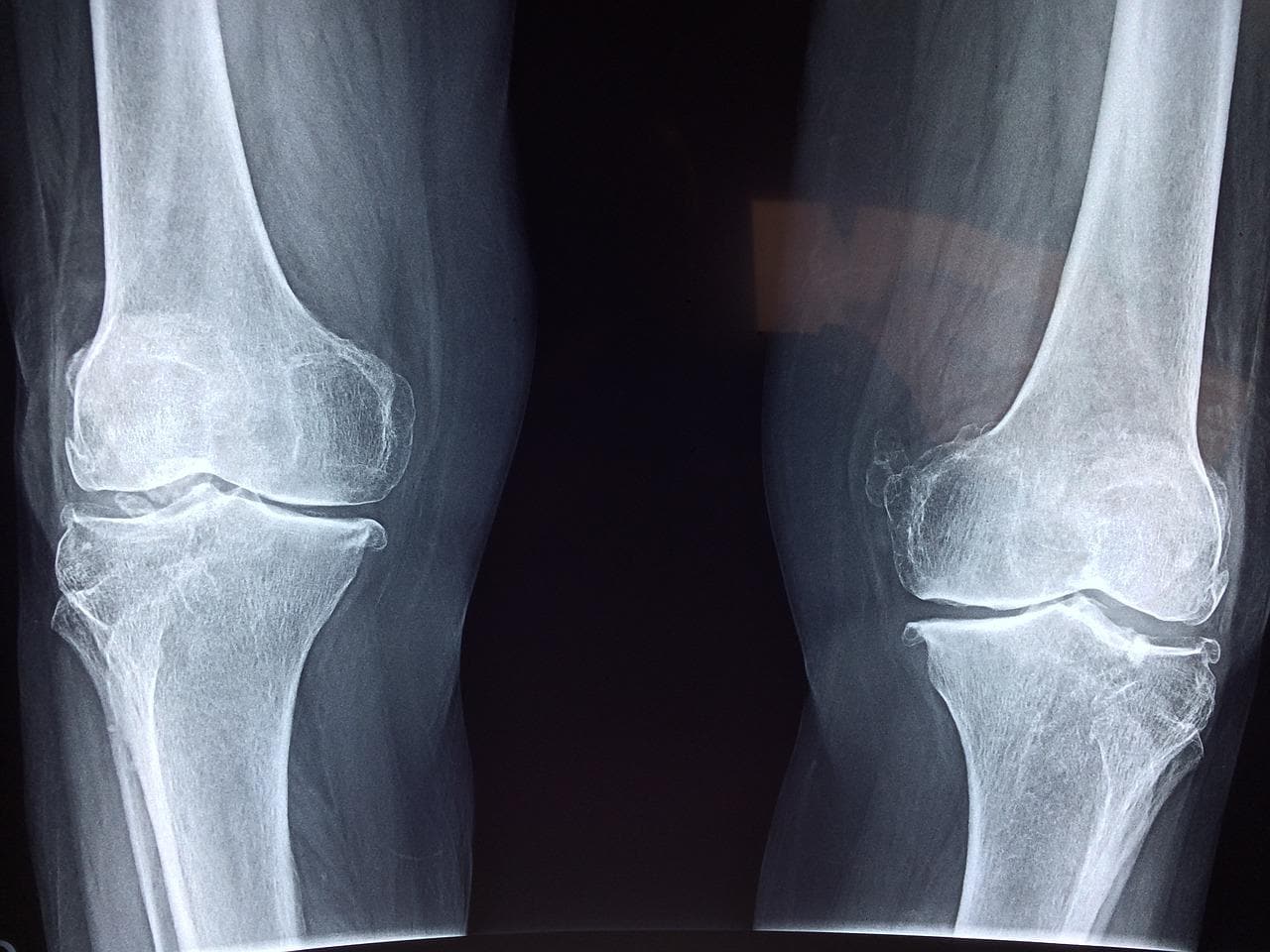
Bone loss is inevitable as we age. Women lose bone mass after menopause. Bone mass decreases as vertebrae lose their mineral content and become thinner. The spinal column becomes curved and compressed as the muscles weaken, and bone spurs develop. Additionally, foot arches become less pronounced, contributing to the slight loss of height. The good news is that preventing osteoporosis is not as difficult as it seems. Fortunately, there are many natural remedies for bone loss and strengthening.
For more information on aging healthy, click here and visit our blog page.